Ebolavirus
Ron Smith, MD
Ebola in the News
(Fred Murphy, artificially colored initial image of Ebola shown)
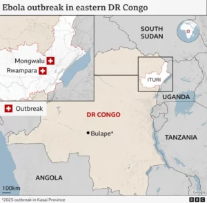 The World Health Organization (WHO) has declared an Ebola outbreak in the Democratic Republic of Congo a public health emergency of international concern. The agency said the outbreak in DR Congo's eastern Ituri province, which has seen around 246 suspected cases and 80 deaths reported, does not meet the criteria of a pandemic emergency.
Quick Overview
History of Ebola
In 1976, Fred Murphy, a virologist working at the CDC, saw the long, filamentous Ebola virus for the first time under electron microscopy and took the artificially colored image on this page. He was also the only expert that had looked at the Marburg virus, a close cousin to Ebola.
Ebola hemorrhagic fever has become know as Ebola Virus Disease, or EVD. Initially a rare disease, it has repeated on even ongoing epidemics in Africa. Various strains of the virus have now been seen around the globe.
It is spread through contact with either an infected wild animal or another person. There is variability of mortality, and in the case of the Reston Ebolavirus, known as RESTV, even symptoms it exhibits. Ebolavirus has an extremely efficient ability to be absorded into living cells, where it replicates explosively. The virus reproduces so quickly that it overwhelms the host immune response. Before antibodies can be produced against the spike proteins that attach to, and cause absorption by, the target cell, widespread cellular infection occurs in all tissues, other than bone.
The initial infection appears to come through wild infected animals which harbor the virus without being harmed by it. In Africa, a staple of the diet in areas where Ebola has been seen seem to begin with consumption of “bush meat” from these animals. The local or rural form gives way to the urban form.
Once infected, the virus spread from person to person. All body fluids team with Ebola. While the incubation period last for up to twenty-one days, the first symptom of fever and headache appears around nine days or so. Historically, death from widespread hemorrhage occurs often by around twelve to fourteen days.
There are different strains of Ebola which exhibit different mortality rates. Ebola Zaire, was named for the Ebola River near the village of Yambuku in what is today known as the Democratic Republic of Congo. The word “Ebola” means black, and the acronym EBOV refers to the Ebola Zaire strain. The mortality rate for Ebola Zaire. Ebola Sudan, in retrospect, was probably the first on the scene in southern Sudan which is near Yambuku. It is designated by the acronym SUDV. EBOV and SUDV have mortality rates typically in the 90% and 50% range, and these two account for the majority of Ebola cases in the world. Mortality rates seems to be affected by the speed of diagnosis and implimentation of intensive supportive care and fluids. This is not a given though, as many notable and well-trained African physicians and other health care workers died after caring for Ebola patients. The overall mortality among Ebola strains is around 50%.
The Ebolavirus strain discovered in Reston, Virginia and Alice, Texas at the Hazelton monkey facilities wreak havoc among monkeys, but cause few, in any symptoms, among humans. The book The Hot Zone and the National Geographic series by the same title together tell a chilling history of Ebola in the US. It is known by the acronym RESTV, and it originated from a monkey farm in the Philippines which sold the animals to the Hazelton company.
The Taï Forest strain, known as TAFV, was isolated in Parc National de Taï, in Côte d’Ivoire. The single known case of TAFV was contracted by a forest researcher who survived the illness.
The Ebola Bombali strain, known as BOMV, was found in Angolan free-tailed bats in Taita Hills, Kenya. Infection in humans was not found in my research.
The chain of infection seems clear though the index animal source of Ebola has yet to be confirmed. Infection brews in wild animals which are consumed by local populations. Person to person spread is quick and efficient through body contact which is especially enhanced with the twenty-one day asymptomatic incubation period. Prodromal symptoms include fever, muscle aches and pain, fatigue, sore throat, and headache, and can easily be deemed a “case of the flu.” Vomiting and diarrhea ensue and become bloody and a hemorrhagic skin rash appears. Bleeding progresses both internally in all organs, and externally from all body orifices. Mucous membrane linings of the gut are sloughed off and expulsed with the diarrhea, and other mucous membranes in the stomach, etc., slough off as well. Liver enzymes skyrocket as vast numbers of hepatic cells rupture and liquefy. Hemorrhage affects the brain and a morbid, blank stare ensues. The kidneys fail as the virus kills renal cells. Severe pain and agony precedes death from multiple organ and circulatory failure. All body fluids teem with billions upon billions of infectious Ebola virus particles.
Cultural practices vary by locale, and have been problematic in Africa where touching and kissing deceased persons at funerals occur. The virus remains viable on the body for a substantial period after death, and the contagion is so easy that even with the strictest of biological barrier protections there is a risk.
Diagnosis
Diagnostic testing can be confirmed with these possible methods. All samples should be handled with level 4 biohazard precautions.
- antibody-capture enzyme-linked immunosorbent assay (ELISA)
- antigen-capture detection tests
- serum neutralization test
- reverse transcriptase polymerase chain reaction (RT-PCR) assay
- electron microscopy
- virus isolation by cell culture.
Treatment
Antiviral treatments so far have not been terribly effective. Intravenous fluids and blood transfusions are important, but once the gut begins to slough its mucosal membrane, these treatments become futile. There are some drug trials you can read about here. Blood from recovered patients has been used in the African field as a last effort to save patients. The monoclonal antibodies Inmazeb and Ebanga are approved by the US Food and Drug Administration.
Vaccines
There are two vaccines that show particular promise, though supplies are probably limited because of the high manufacturing cost compared to the ability to cover research expenses. Ervebo was approved in December 2020 by the US Food and Drug Administration. The WHO prequalified its use for Ebola Zaire, EBOV, in those 18 and older who are not pregnant or nursing. In the 2018-2020 Ebola outbreak in the Congo it was given under compassionate using it to “ring” a wall of immune individuals around outbreak areas.
The second vaccine is a really a combination of the Ervebo vaccine given first followed 8 weeks later by a subsequent dose of the newer Mvabea vaccine. This combination can be given to those 1 year and older, but because of the long time frame between doses, is not useful in epidemic outbreaks.
Considerations for Survivors
Recovery from Ebola virus does not mean all is necessarily well. Ebola can persist in semen, testicular tissue, eyeball fluids, breast milk, and pregnancy related fluids. Relapse is not common, but not unknown either, and the reasons for it are not understood. The PCR, or polymerase chain reaction, can be persistent positive for even up to nine months after recovery from acute disease. The reasons are not know, but I must point out that PCR testing is a screening test and cannot be used as a definitive diagnostic.
Ebolavirus Vaccine History
Prior to the 2014 outbreaks of Ebola in west Africa, Ebolavirus disease (EVD or EHF) outbreaks were rural, and through aggressive contact tracing were effectively contained and ended. The source host for this filovirus was then and continues to be unknown.
Then in 2014, the virus got into the urban areas of several west African countries. The epidemic was traced back to an eighteen-month-old boy in Guniea. At that time, Médecins Sans Frontières and Samaritan’s Purse were the only two organizations responding clinically on the ground in the Liberia, Guinea, and Sierra Leone areas. The outbreak was much larger than any before since previous occurrences were rural and not urban. Médecins Sans Frontières and Samaritan’s Purse were taxed to the breaking point and the outbreak in Guinea, Sierra Leone, and Liberia became the largest in history dwarfing all previous ones.
The development of the Ebola vaccine is far from recent and really began in the mid 1990s. Prior that time, vaccines were based on sub-units of the infectious agent. That is, virus or bacteria had to be grown, harvested and only the “active” proteins retrieved which were included in the vaccine. The earlier lessons of the Cutter Incident with the polio vaccine and the high frequency of side effects with the whole cell pertussis vaccine demonstrated the need for a more targeted approach.
In the mid-1990s, Katalin Karikó, a Hungarian-born scientist, pioneered a new approach using mRNA instead of sub-units. Rather than collecting proteins from viruses and bacteria grown in living tissues, her idea was to develop a vaccine using the specific mRNA which coded very specifically for things like the spikes on viruses which attached them to the host cell membrane which then engulfed and “swallowed” the virion. If antibodies to these spike proteins were present already, then virus particles could never attach and never be engulfed by the host cell.
Karikó’s task was to somehow get the mRNA that coded for these spikes into the host cell whereby only the spikes would be produced. Once presented to the immune system, the proper antibody would be made and memorized. Future challenges by the actual virus then would be met swiftly with antibodies attacking the spike proteins preventing infection of the host cell. The University of Wisconsin had already proved this technique worked in mice in 1990.
The problem of creating mRNA was the main issue. Genomic sequencing became partner to all this as sequencing wholesale DNA and RNA strands became possible and very quick. It took only about 48 hours to sequence the mRNA for SARS-CoV-2 in 2019. A second problem with mRNA is how quickly it is degraded and disposed of outside the host cell. A virion with the spike proteins easily carried that mRNA code into the host cell, but simply injecting mRNA into a person won’t do the same.
With the 2014 Ebola outbreak, vaccine development first started using a viral vector to get the mRNA into the host. The idea is to attach the target mRNA to that of another virus which is not lethal and mostly innocuous. That virus would then be the vector by which the host cell would then produce the target spike protein that would in turn lead to antibody production against Ebolavirus.
In the 1990s, John “jack” Rose at Yale used a livestock virus called VSV (vesicular stomatitis virus) as this viral vector. Heinz Feldmann was instrumental in fusing the key Ebola mRNA onto VSV at the Philipps-Universität Marburg in Germany. This vaccine was called rVSV-ZEBOV. He tested it in animals before leaving the Canada Nation Microbiology Laboratory in Winnipeg in 2008. Steven Jones, an associate of his whose name is on the Ebola vaccine patent, began development of human-grade vaccine for the initial testing trials.
As the 2014 Ebola epidemic unfolded, Gary Kobinger, the head of special pathogesn at the National Laboratory in Winnipeg, was watching. His team had been working on the Ebola vaccine for years which showed great promise in animal testing. Vaccine development had gone so far as to produce one ready for human trials. The WHO declined his offer of the vaccine, however, and probably out of Africa’s history of unethical vaccine development by western entities years earlier with polio.
Essentially as a result of the 2014 Ebola outbreak, Merck finally picked up rVSV-ZEBOV, and the first viral vector vaccine for Ebola was branded as Ervebo. The VSV virus vector would go on into research for flu, mealses, SARS, and ZIKA vaccines. The vaccine was initially approved for compassionate use in 2018. It proved highly effective, and was FDA approved in 2019.
In the 2014 epidemic, both Dr. Kent Brantly and Nancy Writebol working for Samaritan’s Purse in Liberia contracted Ebola Zaire (Facing Darkness). This was, of course, before the Ervebo vaccine was available. However, another technology called ZMapp was already in testing and was available for experimental use.
ZMapp developed by Mapp Biopharmaceutical is not a vaccine, but rather three plant-made monoclonal antibodies which attack the Ebola virus. The tobacco plant Nicotiana benthamiana is infected with a viral vector. There are a number of plant viruses that are effective vectors such as tobamoviruses, Potexviruses (e.g. Potato Virus X) , Tobraviruses (e.g., tobacco rattle virus), Geminiviruses (e.g., bean yellow dwarf virus), and Comoviruses (e.g. cowpea mosaic virus).
ZMapp was effective in saving 100% of rhesus macaques infected with Ebola even up to 5 days before administration. Many animals already showed symptoms and had Ebola-driven laboratory aberrations before the drug. The drug was given to both Dr. Brantly and Writebol. They both survived Ebola Zaire. Miguel Pajares, a Spanish priest, received the third course of ZMapp but subsequently died from Ebola.
Viral vector vaccines led to the development of newer mRNA vaccines which have a lipid coating. This coating similarly acts like a virion so that the virus spike protein is manufactured from the mRNA and then released without harming the host cell. The immune cells then process the spike protein and create antibodies to it. Free of the overwhelming assault of viruses like Ebola and SARS-CoV-2, the immune system is equipped to fight the infection without ever having been infected with the pathogen. mRNA vaccines may well signal newer, safer, and more effective measures than many current vaccines.1
 Copyright secured by Digiprove © 2021 Ronnie Smith
ENDNOTES
- Historical information sources: ‘Against all odds’: The inside story of how scientists across three continents produced an Ebola vaccine, ZMapp, ↩
Table of Ebola (EBV) Strains.
Kingdom: Riboviria, Phylum: Orthornavirae, Class: Negarnaviricota, Order: Monjiviricetes, Order: Mononegavirales, Family: Filoviridae, Genus: Ebolavirus, Species: (as shown below)
Scrolling Table of Current & Past Occurrences of Ebola (EBV) Infection.
Source: WHO Ebola virus disease
How To Use The Ebolavirus Map.
There well over a hundred map markers on the map. When you do a search, all available information for each marker is compared to your search criteria. If you search for “Congo” then the search will return both marker headings and descriptions that match. If you want to search only the marker labels, then use “Congo;” instead. If you want to search for a particular strain of virus, then use the correct acronym. If you want to see outbreaks in a ceration year, then you can use “1976” or “1976.” to get the correct results.
Copyright secured by Digiprove © 2021 Ronnie Smith