Kyasanur Forest Disease

Ron Smith, MD

Kyasanur Forest Disease (KFD) in the News

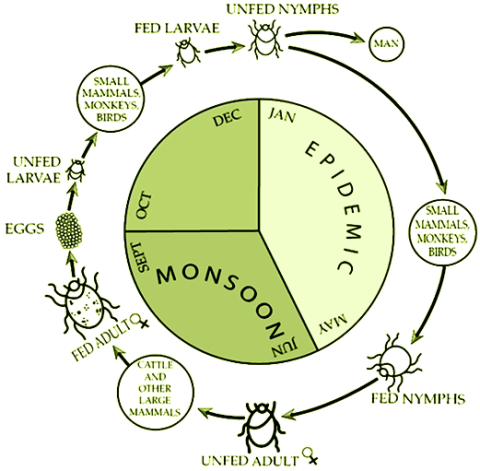
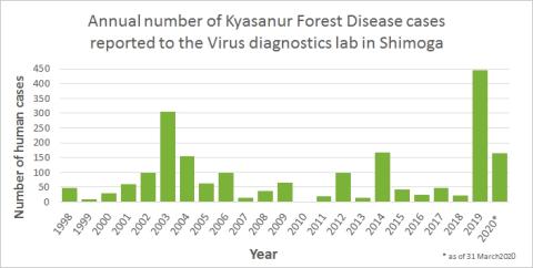
CDC. Kyasanur Forest disease (KFD) is caused by Kyasanur Forest disease virus (KFDV), a member of the virus family Flaviviridae. KFDV was identified in 1957 when it was isolated from a sick monkey from the Kyasanur Forest in Karnataka (formerly Mysore) State, India. Since then, between 400-500 humans cases per year have been reported. Hard ticks (Hemaphysalis spinigera) are the reservoir of KFD virus and once infected, remain so for life. Rodents, shrews, and monkeys are common hosts for KFDV after being bitten by an infected tick. KFDV can cause epizootics with high fatality in primates, but typically mortality is around 5%.

.


Saturday, 1 January 2033

What is Kyanasur Forest Disease (aka KFD or Monkey Fever)?

KFD, also referred to as Monkey Fever, is a tick-borne viral haemorrhagic disease, which can be fatal to humans and other primates. The causal agent, Kyasanur Forest Disease Virus (family Flaviviridae, genus Flavivirus), is a member of the tick-borne encephalitis (TBE) complex. It is transmitted by a range of tick species, with Haemophysalis spinigera being considered the principal vector.


Thursday, 16 March 2023

Why areca nut plantations are driving 'monkey fever'

A tick-borne infection often spread in areca nut plantations in India is among several on the rise around the world. Can we get better at tackling these outbreaks?


Quick Overview

Crimean-Congo Hemorrhagic Fever


Crimean-Congo hemorrhagic fever (CCHF) is caused by infection with a tick-borne virus (Nairovirus) in the family Bunyaviridae. The disease was first characterized in the Crimea in 1944 and given the name Crimean hemorrhagic fever. It was then later recognized in 1969 as the cause of illness in the Congo, thus resulting in the current name of the disease.

Crimean-Congo hemorrhagic fever is found in Eastern Europe, particularly in the former Soviet Union, throughout the Mediterranean, in northwestern China, central Asia, southern Europe, Africa, the Middle East, and the Indian subcontinent.

Transmission

Ixodid (hard) ticks, especially those of the genus, Hyalomma, are both a reservoir and a vector for the CCHF virus. Numerous wild and domestic animals, such as cattle, goats, sheep and hares, serve as amplifying hosts for the virus. Transmission to humans occurs through contact with infected ticks or animal blood. CCHF can be transmitted from one infected human to another by contact with infectious blood or body fluids. Documented spread of CCHF has also occurred in hospitals due to improper sterilization of medical equipment, reuse of injection needles, and contamination of medical supplies.

Signs and Symptoms

The onset of CCHF is sudden, with initial signs and symptoms including headache, high fever, back pain, joint pain, stomach pain, and vomiting. Red eyes, a flushed face, a red throat, and petechiae (red spots) on the palate are common. Symptoms may also include jaundice, and in severe cases, changes in mood and sensory perception.

As the illness progresses, large areas of severe bruising, severe nosebleeds, and uncontrolled bleeding at injection sites can be seen, beginning on about the fourth day of illness and lasting for about two weeks. In documented outbreaks of CCHF, fatality rates in hospitalized patients have ranged from 9% to as high as 50%.

The long-term effects of CCHF infection have not been studied well enough in survivors to determine whether or not specific complications exist. However, recovery is slow.

Risk of Exposure

Animal herders, livestock workers, and slaughterhouse workers in endemic areas are at risk of CCHF. Healthcare workers in endemic areas are at risk of infection through unprotected contact with infectious blood and body fluids. Individuals and international travelers with contact to livestock in endemic regions may also be exposed.

Diagnosis

Laboratory tests that are used to diagnose CCHF include antigen-capture enzyme-linked immunosorbent assay (ELISA), real time polymerase chain reaction (RT-PCR), virus isolation attempts, and detection of antibody by ELISA (IgG and IgM). Laboratory diagnosis of a patient with a clinical history compatible with CCHF can be made during the acute phase of the disease by using the combination of detection of the viral antigen (ELISA antigen capture), viral RNA sequence (RT-PCR) in the blood or in tissues collected from a fatal case and virus isolation. Immunohistochemical staining can also show evidence of viral antigen in formalin-fixed tissues. Later in the course of the disease, in people surviving, antibodies can be found in the blood. But antigen, viral RNA and virus are no more present and detectable.

Treatment

Treatment for CCHF is primarily supportive. Care should include careful attention to fluid balance and correction of electrolyte abnormalities, oxygenation and hemodynamic support, and appropriate treatment of secondary infections. The virus is sensitive in vitro to the antiviral drug ribavirin. It has been used in the treatment of CCHF patients reportedly with some benefit.

Recovery

The long-term effects of CCHF infection have not been studied well enough in survivors to determine whether or not specific complications exist. However, recovery is slow.

Prevention

Agricultural workers and others working with animals should use insect repellent on exposed skin and clothing. Insect repellants containing DEET (N, N-diethyl-m-toluamide) are the most effective in warding off ticks. Wearing gloves and other protective clothing is recommended. Individuals should also avoid contact with the blood and body fluids of livestock or humans who show symptoms of infection. It is important for healthcare workers to use proper infection control precautions to prevent occupational exposure.

Resources

Khan A, et al. Viral Hemorrhagic Fevers. Seminars in Pediatric Infectious Diseases. Philadelphia: WB Saunders Co., 1997;8 (suppl 1):64-73. Peters CJ. Viral Hemorrhagic Fevers. Viral Pathogenesis. New York: Lippincott-Raven Publishers, 1997:779-794.

Khan AS, et al. Viral Hemorrhagic Fevers and Hantavirus Pulmonary Syndrome. In HF Conn, RH Clohecy, RB Conn, eds. Current Diagnosis 9. Philadelphia: WB Saunders Co., 1997:193-194..

After an incubation period of 3-8 days, the symptoms of KFD begin suddenly with chills, fever, and headache. Severe muscle pain with vomiting, gastrointestinal symptoms and bleeding problems may occur 3-4 days after initial symptom onset. Patients may experience abnormally low blood pressure, and low platelet, red blood cell, and white blood cell counts.

After 1-2 weeks of symptoms, some patients recover without complication. However, the illness is biphasic for a subset of patients (10-20%) who experience a second wave of symptoms at the beginning of the third week. These symptoms include fever and signs of neurological manifestations, such as severe headache, mental disturbances, tremors, and vision deficits.

The estimated case-fatality rate is from 3 to 5% for KFD.

Diagnosis can be made in the early stage of illness by molecular detection by PCR or virus isolation from blood. Later, serologic testing using enzyme-linked immunosorbent serologic assay (ELISA) can be performed.